Dr James 'Mac' Shine

Dr Mac Shine, Robinson/SOAR Fellow, The University of Sydney.

Mac is a theoretical neuroscientist working to understand the mechanisms of cognition and attention using functional brain imaging, both in health and disease. He is currently working as a Robinson/SOAR fellow at The University of Sydney to understand the factors that drive the network-level reorganization of the human brain. He completed my post-doc with Russell Poldrack at Stanford University, received his PhD from Sydney University in 2013 (PI: Simon JG Lewis), his MBBS from Sydney University in 2007 and his BSc from the University of Sydney in 2003.

Forefront Group:

  • Brain and Mind Centre Functional Magnetic Resonance Imaging Research Group Leader

Affiliate Organisations

https://www.forefrontresearch.org/wp-admin/media-upload.php?post_id=1312&type=image&TB_iframe=1

The University of Sydney, Sydney Medical School, Center for Complex Systems

Expertise:

  • Functional neuroimaging
  • Computational modelling
  • Systems neuroscience

Neurodegeneration of interest:

PD, DLB, AD

Specific Skills:

  • Medical Doctor
  • Functional Neuroimaging
  • Cognitive Neuroscience
  • Computational Modelling
  • Evolutionary Neuroscience

Project - Noradrenaline and Cognitive Dysfunction - Interrogating the Neglected Symptoms of Parkinson’s disease

Research Project Abstract

The first stage of our proposal uses a suite of innovative neuroimaging methods in combination with peripheral markers of the noradrenergic system. We apply these tools to a cohort of healthy participants as they undergo functional MRI while performing diverse cognitive tasks. In doing so, Aim I will quantify relationships between ascending sympathetic tone and cognitive function in a cohort of healthy individuals. These experiments will establish mechanistic links between cognitive function, network architecture and the ascending arousal system.

A similar multi-modal imaging approach will then be used to characterize the central and autonomic neural basis of cognitive dysfunction in Parkinson’s disease. Aim II will examine the relationship between cognitive impairment and deficits in autonomic arousal in individuals with Parkinson’s disease. Specifically, we will use multiple autonomic biomarkers to confirm the presence of central noradrenergic deficits across a diverse cohort of individuals with Parkinson’s disease. We will then assess the multimodal neuroimaging signature of cognitive deficits in Parkinson’s disease, testing the hypothesis that an abnormal arousal system predisposes individuals to impaired cognitive function in Parkinson’s disease through a failure to effectively integrate the network architecture of the brain during cognitive tasks.

Project tag with a disease

PD, DLB

Challenges within the field

  • Imaging the ascending arousal system, which has a small locus, wide influence and intimate relationships with sources of systemic noise.
  • Conceptualising cognitive function in a way that is commensurate with the models we use to understand the impact of the ascending arousal system on the rest of the brain.

Research Project Description

Structural markers of noradrenergic impairment – recent advances in structural imaging have led to the development of novel sequences that can provide structural indices of noradrenergic cell integrity. T1-neuromelanin scans are sensitive to concentrations of neuromelanin, which is particularly high in the noradrenergic locus coeruleus; and magnetic resonance spectroscopy of the pons identifies the strength of chemical signatures of particular neurotransmitters in specific locations in the brain. Previous work has demonstrated a positive link between neuromelanin intensity and whole-brain noradrenergic uptake (2). We predict similar relationships between structural noradrenergic integrity and pupil diameter, both during rest and as a function of task performance. Recent work from other groups (2) suggests that this experiment has a high chance of success.

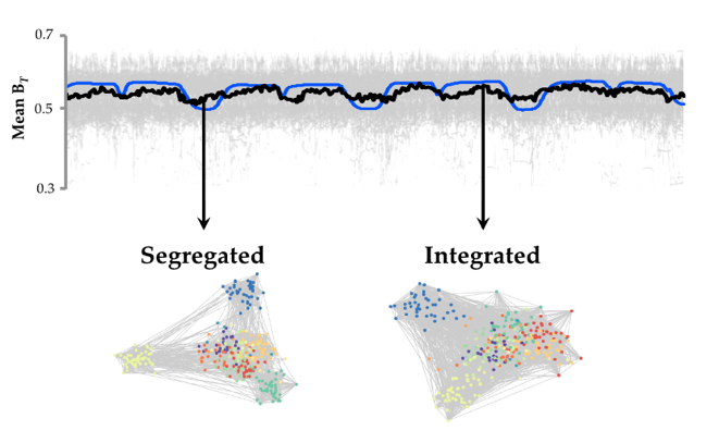
Noradrenergic network signatures during the resting state – We will use our recently devised pipeline for analyzing fluctuations in network structure over time (16) to track alterations in functional network structure while individuals with Parkinson’s disease perform the suite of cognitive tasks. In previous work, we demonstrated that the functional network signature of the brain fluctuates over time between states of high and low ‘integration’ (16). In addition, we documented a relationship between fluctuations in pupil diameter (a measure of central noradrenergic tone) and integration within a network of frontoparietal, striatal and thalamic regions. Given the positive relationship between network structure and pupil diameter, we hypothesize that individuals with a dysfunctional central noradrenergic system (such as those with Parkinson’s disease and cognitive impairment) will demonstrate abnormalities in patterns of network architecture that are sensitive to noradrenergic mechanisms – namely, brain-wide integration. To test this prediction, we will investigate the relationship between network structure and network fluctuations during the ‘resting state’.

Noradrenergic network signatures during performance of cognitive tasks – To determine whether abnormalities in our measures of central sympathetic tone relate to ongoing deficits in cognition, we will investigate fluctuations in network topology during the performance of a suite of cognitively challenging tasks. These tasks will include an ‘N-back’ task (http://www.cognitiveatlas.org/) to investigate working memory and cognitive control; a Stop-Signal task to investigate response inhibition; and a ‘global-local’ task to investigate attentional focus and the ability to avoid distraction. Each of these domains is impaired in Parkinson’s disease (59) and is also to be sensitive to noradrenergic function (60-62).

Research Objectives

Brain mechanisms: There is widespread consensus that identifying the neural processes underlying neurodegenerative disease is pivotal to advancing better treatments, as these processes specify the mechanisms (e.g. direct, psychological, pharmacological) that can be modulated to confer potential benefits to people with the disorders. Indeed, understanding the basics of brain-behaviour processes is currently the Number One strategic priority of the NIMH (Strategic Plan, 2008).

Treatment: Impairments in cognition pervade subjects’ lives and are often highly distressing, leading to high morbidity with nursing home placement (4). If our experiments provide strong support for our hypothesis, we can immediately identify a suite of novel therapeutic targets, including: a) increasing synaptic noradrenaline by blocking reuptake (63); or b) increasing noradrenaline synthesis (64). In addition, the results of Aim 1 may also validate an inexpensive measure for tracking alterations in cognitive function in the clinical setting.

Reverse Translation: There are many ways in which the noradrenergic system can be causally manipulated in animal models (such as the mouse), using tools such as optogenetics to drive the system towards improved or impaired cognitive performance. That power provides opportunities to uncover nuanced ways to improve cognitive function through the enhancement of the interaction between the sympathetic and central nervous systems.

Other disorders: Parkinson’s disease is not the only neurodegenerative disorder associated with pathological involvement of the noradrenergic system. Indeed, pathology of the locus coeruleus may be an indicator of cognitive deficits in Alzheimer’s disease (65). Hence, our work may provide a template for researchers in other areas of clinical neuroscience.

Key Publications from this project

  • Shine, J.M., Bissett, P.G., Bell, P.T., Koyejo, O., Balsters, J.H., Gorgolewski, K.J., Moodie, C.A. and Poldrack, R.A. (2016). The Dynamics of Functional Brain Networks: Integrated Network States during Cognitive Task Performance. Neuron, 92(2):544-554. (PMID: 27693256).
    Cited 240 times (Google Scholar; GS); IF 15.8.
    This study was one of the first to track the network structure of the brain over time using whole-brain functional neuroimaging analyses. We found that the network structure of the brain was highly dynamic, and that fluctuations in the architecture were associated with fast, effective performance on a challenging cognitive task. This work has since been replicated by multiple groups, and forms the basis of a new vantage point on whole-brain dynamics in cognition.
  • Shine, J.M., Koyejo, O., Poldrack, R.A. (2016). Temporal metastates are associated with differential patterns of time-resolved connectivity, network topology, and attention. Proceedings of the National Academy of Sciences, 113(35):9888-91. (PMID: 27528672).
    Cited 105 times (GS); IF 9.6.
    Here, we leveraged a unique single-subject, longitudinal dataset to interrogate the stability of the brain’s network structure over the course of weeks-to-months. We found remarkable flexibility in the brain’s network architecture over time, and further observed that these fluctuations coincided with alterations in self-reported attentional focus and engagement. These ideas have important implications for neuropsychiatric and neurodegenerative diseases in which symptomatology fluctuates over time.
  • Shine, J.M., Breakspear, M., Bell, P.T., Martens, K.A.E., Shine, R., Koyejo, O., Sporns, O. and Poldrack, R.A. (2019). Human cognition involves the dynamic integration of neural activity and neuromodulatory systems. Nature Neuroscience, 22(2), 289-296 (PMID: 30664771).
    Cited 57 times (GS); IF 21.1.
    Here we investigated the spatial, dynamic, and molecular signatures of system-wide neural activity across a range of cognitive tasks. We found that neuronal activity converged onto a low-dimensional manifold that facilitates the execution of diverse task states. Flow within this attractor space was associated with dissociable cognitive functions, unique patterns of network-level topology, and individual differences in fluid intelligence. The axes of the low-dimensional neurocognitive architecture aligned with regional differences in the density of neuromodulatory receptors, which in turn relate to distinct signatures of network controllability estimated from the structural connectome.

Infographic / Medical Diagram / Scientific Diagram / Picture

Key Findings

  • Within the brain, a relatively fixed structural scaffold gives rise to a highly dynamic functional landscape, in which the emergence of momentary neural coalitions forms the basis for complex cognitive functions, learning and conscious experience. This view of brain function highlights the role of individual brain regions within the context of a broader neural network. How these architectures change over time is crucially important for understanding human behavior, which is inherently dynamic. We have recently devised a robust and reproducible pipeline for analyzing fluctuations in network structure over time. We first estimate changes in inter-regional coordination within short temporal windows using an approach designed by CI-Shine and AI-Poldrack. We then slide those windows over time and apply graph-theoretical tools to the resultant covariance matrices. This yields a dynamic estimate of the extent to which the brain is organized into tight-knit communities of regions (also known as ‘modularity’) over the course of minutes (using a weighted- and signed-version of the Louvain algorithm; γ = 1). Using this information, we can then estimate how much each region ‘participates’ across multiple tight-knit communities – a sensitive marker for how ‘integrated’ the brain is at any given point in time. We have shown that an integrated network architecture is associated with fast, effective cognitive performance– see Figure (blue: task block; black: brain network integration).
  • Parkinson’s disease is primarily characterized by diminished dopaminergic function; however, the impact of these impairments on large-scale brain dynamics remains unclear. It has been difficult to disentangle the direct effects of Parkinson’s disease from compensatory changes that reconfigure the functional signature of the whole brain network. To examine the causal role of dopamine depletion in network-level topology, we investigated time-varying network structure in 37 individuals with idiopathic Parkinson’s disease, both ON and OFF dopamine replacement therapy, along with 50 age-matched, healthy control subjects using resting state functional MRI. By tracking dynamic network-level topology, we found that the Parkinson’s disease OFF state was associated with greater network-level integration than in the ON state. The extent of integration in the OFF state inversely correlated with motor symptom severity, suggesting that a shift toward a more integrated network topology may be a compensatory mechanism associated with preserved motor function in the dopamine depleted OFF state. Furthermore, we were able to demonstrate that measures of both cognitive and brain reserve (i.e. premorbid intelligence and whole brain grey matter volume) had a positive relationship with the relative increase in network integration observed in the dopaminergic OFF state. This suggests that each of these factors plays an important role in promoting network integration in the dopaminergic OFF state. Our findings provide a mechanistic basis for understanding the Parkinson’s disease OFF state and provide a further conceptual link with network-level reconfiguration. Together, our results highlight the mechanisms responsible for pathological and compensatory change in Parkinson’s disease.